Minggu, 29 Agustus 2010

Tindakan percutaneus transhepatic biliary drainage

PTBD (PERCUTANEUS TRANSHEPATIC BILIARY DRAINAGE)

Penyebab utama perforasi duktus biliaris yaitu komplikasi tindakan laparoscopi cholecystectomi atau enterobiliaris anastomis. Penanganan penderita tersebut dengan pembedahan atau endoscopic biliary drainage. Penanganan tersebut kadang tidak berhasil utamanya jika defek duktus biliaris post operasi yang besar atau perforasi biliary oleh karena acute necrotizing pancreatitis berat. Alternatif tindakan PTBD, dalam beberapa laporan kasus penelitian, yang berhasil mengatasi obstruksi biliaris dengan PTC (percutaneus transhepatic cholangiography) diikuti penempatan kateter untuk drainase empedu (PTBD) dilakukan pada penderita obstruksi biliary oleh karena tumor ganas yang unresectable, sclerosing kolangitis, striktur pasca operasi atau transplantasi hepar.

Indikasi PTBD :

  1. Obstruksi dari ductus biliaris, sehingga dapat diketahui level, seberapa berat obstruksi dan penyebab obstruksi

  2. Sebagai bentuk persiapan preoperatif, yang dapat membantu surgery mengetahui permasalahan duktus biliaris

  3. Trauma pada duktus biliaris

  4. Sebagai jalan untuk pemasangan internal stent

  5. Penderita dengan infeksi duktus biliaris (Cholangitis)

Tindakan PTBD (percutaneus transhepatic biliary drainage) yaitu suatu manajemen tindakan dekompressi penderita obstruksi biliaris dengan prosedur menempatkan jarum kecil dari luar kulit bagian upper abdominal dextra. Jarum kecil ditusukkan menuju ke hepar dan dengan bantuan fluoroscopi serta kontras ditempatkan pada duktus biliaris, jarum kemudian digantikan oleh guiding wire (penuntun kateter) dan kateter atau tube (berfungsi sebagai drainage) yang fleksibel.

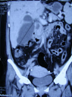
Contoh kasus RSUD Dr. Soetoma, Surabaya

Tt/39 th/ Pr/ Keluhan utama : ikterus dan nyeri upper abdominal dextra, post operasi cholecystectomi ec. Rest stone DD stricture 1 minggu


Lab : Bilirubin direct 14,98 mg/dl, bilirubin total 18,14 mg/dl, SGOT 120 u/L, SGPT 119 u/L, albumin 3,6 g/dl, alkali phosphatase 1699, kreatinin serum 1,23 mg/dl, kolesterol total 532 mg/dl, trigliserida 337 mg/dl


Kesan : Pelebaran intrahepatik dan ekstrahepatik duktus biliaris ec. obstruksi duktus biliaris setinggi ampula vateri, disertai pocketed duktus biliaris ukuran 7,8x6,8x10,2 cm3 (Biloma post operasi cholecystectomi?)

Usul tindakan : dekompressi (PTBD)

Abd. Haris


Jumat, 20 Agustus 2010

Carotid Cavernous Fistula

Carotid cavernous fistula merupakan kelainan yang umumnya karena traumatik pada kepala atau wajah dengan gambaran klinis yang khas, kejadian akut dan progressif penurunan penglihatan, proptosis, chemosis dan diplopia. Diagnosis ditegakkan berdasarkan pemeriksaan CTA (computed tomography angiography), MRA (magnetik resonansi angiography) atau arteriography arteri carotis. Kelainan tersebut terjadi karena hubungan atau fistulasi antara arteri carotis interna atau externa dan sinus cavernous.

CCF ini terbagi atas beberapa tipe :

  • Tipe-A fistula berasal langsung dari a carotis interna dengan sinus cavernosus (direct)

  • Tipe-B fistula berasal dari cabang meningeal dari a carotis interna dengan sinus cavernosus (indirect)

  • Tipe-C fistula berasal dari dari cabang meningeal dari a. carotis externa dengan sinus cavernosus (indirect)

  • Tipe-D fistula berasal dari cabang meningeal a. carotis interna dan a. carotis externa dengan sinus cavernosus (indirect)

Arteriogram penting dalam menentukan lokasi yang tepat dari fistula, suplai arteri, dan pola drain vena. Arteriography juga menyediakan akses untuk pengobatan definitif dari CCF. Saat ini, cara yang paling baik untuk mengobati CCF adalah melalui rute endovascular.

TINDAKAN

Tipe fistula A dapat ditindaki dengan endovaskular embolisasi pada fistula dengan menggunakan detachable ballon, posisi detachable ballon untuk mengoklusi fistula dan mempertahankan patensi dari arteri carotis interna. Keadaan pembuluh darah vena yang mengalir ke jugular interna dan sinus petrosal tidak lagi mendapat akses dari fistula tapi dari sinus cavernosus sendiri

Tipe fistula B, C dan D yang mempunyai fistula kecil sehingga dengan memberikan tekanan sendiri pada arteri carotis 20-30 detik 4 kali perjam untuk menimbulkan trombosis pada fistula. Penderita di instruksikan menekan a. carotis communis sisi yang sakit (ipsilateral) dengan tangan (kontralateral) dan jangan sampai terjadi iskemia selama penekanan. Jika kompresi ini tidak efektif dapat dilakukan selective endovascular embolization pada fistula arteri carotis externa. Pilihan material embolik yang available yaitu polyvinyl alcohol

Contoh : Kasus traumatik cedera kepala ringan, RSU Dr. Soetomo yang telah dilakukan Pemeriksaan arteriography dan CT angiography :


Kesimpulan kasus : Direct (Type A) carotid cavernous fistula (arteri carotis interna sinistra dengan sinus cavernous)

Rencana tindakan : Embolisasi fistula dengan detachable ballon

Rujukan :

Kobayashi N et. al ; endovascular treatment strategy for direct CCF resulting from rupture of intracavernous carotid aneurysm; AJRN Am J Neuroradiol 24:1789-1786, oktober 2003

Koenigsberg R et.al; Carotid-Cavernous Fistula Imaging; emedicine.medscape.com,update 2009

(Dina, Cristine, Haris)


Rabu, 18 Agustus 2010

terapi pada carotid cavernous fistula

 Indications for treatment of CCF
       - glaucoma
       - diplopia
       - intolerable bruit or headache
       - severe proptosis (causing exposure keratopathy)
Type of treatment :
        - Surgical treatment 
            (include ligation of the external & internal carotid arteries)
        - Fistula embolization
             (with particles, glue, detachable ballons, & thrombogenic microcoils)
Direct fistulas are best treated with a detachable ballon.
Treatment of cavernous dural arteriovenous fistulas is usually done using a trans-arterial approach, however in many complicated cases may not unsuccesful, but can be treated with trans-venous embolization.
Complications of treatment may due to the procedure or reopening of the fistula :
        - ophthalmoplegia
        - cerebral infarction
        - ophthalmic artery occlusion
        - increasid proptosis
        - elevated of IOP
        - choroidal detachment
        - venous stasis retinopathy.


CAROTID-CAVERNOUS FISTULATION

= Abnormal communications between the carotid arterial system and the venous cavernous sinus
Broadly classified as either direct or indirect, on the basis of anatomic features depicted on angiograms

Symptomatic direct CCFs (type A):
 - Spontaneously resolve only in rare cases
 - Almost always require urgent treatment, goal: to eliminate flow through the fistula but also to maintain internal carotid patency

Angiography, computed tomography (CT) scanning, magnetic resonance imaging (MRI), and magnetic resonance angiography (MRA) are also useful in assessing the effectiveness of treatment

CT and MRI are the preferred radiologic modalities compared with angiography:
 - Lower incidence of complications
 - Ability to depict peripheral pathologies associated with CCFs (eg, enlargement of cavernous sinus and the ophthalmic vein)
Angiography is used to confirm CT or MRI findings prior to treatment

Plain radiographic findings are most useful for follow-up after embolization therapy, to evaluate balloon positioning or possible leakage


CT finding:

Enlargement of the ipsilateral cavernous sinus
Enlargement and tortuosity of the superior ophthalmic vein
Enlargement of the extraocular muscles
Proptosis

MRI finding:

= CT
Abnormal flow voids in the affected cavernous sinus 
Decreased MRI signal in the involved cavernous sinus
Dilated intercavernous sinuses and intercavernous vessels
Lateral wall convexity of the cavernous sinus
Dilated superior ophthalmic vein, ipsilateral or contralateral
Orbital edema

USG:

Orbital sonograms demonstrate signs similar to those on CT scans and MRIs. In addition, orbital sonogram may demonstrate a reversal of flow direction in the superior ophthalmic vein.
Dilated tortuous veins may be prominent on B-scan echograms. With the A-scan method, dilated ophthalmic veins may be evident. A-scan ultrasonography also can show thickening of the optic nerve.

Nuclear Medicine:

Radionuclide cerebral angiography performed with technetium-99m pertechnetate shows increased uptake of the tracer in the area of the carotid siphons, with rapid clearance. This study is useful in the early postoperative period in a patient with a large CCF repair when angiography may be dangerous.

Angiography:

To accurately identify a carotid-cavernous fistula, selective catheterization of the right and left external and internal carotid arteries and the vertebral arteries is necessary.
Including the entire skull in lateral projection imaging is important.

On an intracavernous carotid arteriogram in a patient with direct CCF, arteriovenous shunting into the cavernous sinus is evident
Immediate filling of the petrosal sinus and/or the ophthalmic vein is commonly evident when the intracavernous carotid artery is injected. Frame rates of greater than 5 frames per second and intracavernous carotid arterial injection rates of greater than 7 mL/s may aid in evaluating the morphology of high-flow fistulas.

The Mehringer-Hieshima maneuver may also be useful in improving delineation of the lesion. This maneuver involves a 2- to 3-mL/s injection into the ipsilateral intracavernous carotid artery with manual compression of the artery below the catheter tip in the neck. This compression allows flow control within the artery to aid in demonstrating the location of the tear.

The Huber maneuver involves an injection of the ipsilateral vertebral artery, with lateral-projection angiography performed by using manual compression of the affected carotid artery during the injection. The retrograde siphon filling of the cavernous sinus is evident. The maneuver helps in identifying the upper extent of the fistula, and it can further help in demonstrating double-hole traumatic fistulas and complete cavernous-intracavernous carotid artery transection.

Kamis, 12 Agustus 2010

pengaruh sistem imun pada terapi hepatoma

PENGARUH SISTEM IMUN PADA TERAPI HEPATOMA

HEPATOMA

= hepatocellular carcinoma
Tu ganas hati primer yg plg sering ditemukan 
E/ : - virus hepatitis B,C,D
  - sirosis hati
  - aflatoksin
  - infeksi
  - keturunan dan ras

Predisposisi
  Pada individu yg tdk imunokompromais :
    respon imun (humoral dan selular) thd  
    kanker .

Efektor sistem imun humoral dan selular pd destruksi kanker

  A. Mekanisme humoral :
      1. Lisis oleh antibodi dan komplemen
      2. Opsonisasi melalui antibodi dan  
          komplemen
      3. Hilangnya adhesi oleh antibodi

  B. Mekanisme selular:
      - destruksi oleh sel CTL/Tc
      - destruksi oleh sel NK
      - destruksi oleh makrofag

Antigen pd kanker :
  - TSA
  - TAA

 Manifestasi paraneoplastik pd hepatoma :

-Hiperkalsemia
-Eritrositosis
-Hiperkolesterolemia
-Alfa feto protein (petanda tu yg baik utk
  hepatoma) ok sel-sel hati mengalami diferensiasi spt pd masa janin.

Antibodi yang dibentuk oleh tubuh terhadap antigen kanker diduga lebih berperan terhadap sel yang bebas ( leukemia, metastase kanker ) dibanding kanker padat. Dengan kata lain destruksi sel kanker lebih efisien bila sel kanker ada dalam suspensi, dan adanya destruksi kanker sulit dibuktikan pada kanker yang padat. Dari sifat-sifat imunologis tersebut diatas maka terapi untuk kanker yang padat termasuk hepatoma adalah lebih sulit dan belum memberikan harapan yang baik.

Mungkin perlu dipertimbangkan  pemberian imunoterapi untuk memanipulasi respons imun terhadap kanker untuk meningkatkan destruksi kanker. Imunoterapi yang dapat diberikan adalah :

    a. Imunoterapi pasif, yaitu :

           - antibodi monoklonal

           - imunotoksin

    b. Imunoterapi aktiv :

           - infus sitokin

     c. Lymphokine activated killer cells

     d. Tumor infiltrating lymphocyte

     e. Macrophage activated killer cells.


Rabu, 11 Agustus 2010

SISTEM IMUN DALAM HUBUNGANNYA DENGAN TERAPI HEPATOMA

SISTEM IMUN DALAM HUBUNGANNYA DENGAN TERAPI HEPATOMA

Wikipedia

  • Virus Hep B/C menginduksi sel2 imun utk menyerang hepatosit sehat secara berulang2

Eur J Gastroenterol.Hepatol. 2004

  • Sel dendritik memproses & mempresentasikan antigen utk mengaktivasi sel T naif & jika dipenuhi antigen tumor, sel dendritik akan menstimulasi respon anti-tumor spesifik yg bersifat tahan lama

The Lancet

  • Aflatoxin menekan imunitas seluler, terutama pd populasi di daerah endemik

Journal of Biomed Science

  • Autofagi:
    Concanavalin A melalui manosa (glikoprotein di membran sel) akan masuk ke mitokondria, menyebabkan autofagi & berakhir dg kematian sel
  • Imunomodulasi:
    Con A (mitogen sel T) mengaktivasi respon imun hepar yg mengeradikasi tumor
    Pembentukan nodul tumor dihambat oleh sel T CD8+ shg diperoleh memori imun thd antigen tumor spesifik


West Indian Med Journal

  • Hepatoma:
    Limfosit T & B meningkat
    Sel NK meningkat
    Makrofag berkurang


World Journal Gastroenterol

  • AFP 20 mg/L menyebabkan ekspresi FasL & TRAIL sel hepatoma meningkat; menghambat ekspresi Fas & TRAILR limfosit
  • Hal ini berakibat sel hepatoma mampu menghindari surveilans imun limfosit host


thefreelibrary

  • AFP:
    Suatu protein antigenik lemah
    Harus dipresentasikan bersamaan dg sel dendritik utk menimbulkan respon imun
    Maka dibuat suatu vaksinasi sel dendritik

Diskusi journal radiologi

PENGARUH TACE ATAU TACI PADA PENDERITA HEPATOCELLULAR CARCINOMA DENGAN GANGGUAN IMUN SISTEM (diskusi 10/08/2010)
1. Pendahuluan
Hepatocelluler carcinoma (HCC) merupakan penyakit kanker terlima terbanyak di dunia, mempunyai prognosis yang buruk sebagai tumor hepar yang maligant. Pada U.S. pasien dengan sirosis oleh karena infeksi kronik hepatitis B atau C, alkoholik dan hemocromatosis mempunyai risiko mendapatkan HCC. Dengan kombinasi studi imaging (Ultrasonography, CT dan MRI) dan elevasi level darah dari alfa-fetoprotein (AFP) menjadi lebih efektif dalam diagnosis. HCC yang resectable (biasanya kecil) dengan fungsi hati yang baik ditindaki dengan reseksi bedah namum yang unresectable biasanya ditindaki dengan TACI (transarterial chemo-infusion) atau TACE (transarterial chemo-embolisasi) sesuai prosedur inklusi maupun eksklusi .
2. Faktor risiko :
a. Hepatitis B atau Hepatitis C
b. Alkohol
c. Alfatoxin B1
d. Drug, medication dan chemical
e. Hemocromatosis
f. Cirrhrosis
3. Pemeriksaan Diagnostik HCC
Sel-sel HCC biasanya memproduksi hormon yg tersebar ke sistem tubuh kemudian memperlihatkan test darah abnormal berupa ;
• High red blood count (erytrocytosis)
• Low blood sugar (hypoglicemia)
• High blood calcium (hypercalcemia)
Alfa feto protein (AFP) mempunyai sensitivitas 60 % dalam menentukan HCC dan 40 % kelainan HCC memperlihatkan nilai normal. Nilai AFP lebih dari 500 ng/4µL dinyatakan sangat mendukung sebagai HCC. AFP yang berkurang berkolerasi dengan ukuran HCC yang juga mengecil sehingga abnormal AFP pada kelainan HCC dapat menjadi marker respon tindakan.
Imaging Study dapat memperlihatkan ukuran, jumlah, keterlibatan pembuluh darah dan penyebaran extrahepatika .
Evaluasi sel-sel hormonal HCC, Alfafetoprotein dan studi imaging pre maupun post tindakan TACE atau TACI dapat disimpulkan adanya komplit atau parsial respon , tidak ada perubahan, atau progressive disease. 
4. Tindakan TACI atau TACE penderita HCC

Tindakan TACI atau TACE sebagai terapi pilihan paliatif. TACI arteri hepatika adalah memberikan dosis tinggi agen kemoterapi langsung ke hepar melalui feeding arteri tumor hepar dan mengurangi komplikasi lokal dari infusi arteri hepatika dengan drug delivery sistem seperti lipiodol, mikrokapsul dan bentuk mikrosphere. TACE arteri hepatika yaitu disamping memberikan agen kemoterapi langsung keorgan target juga dilakukan tindakan embolisasi. Tindakan ini lebih menguntungkan dibanding ligasi arteri hepatika karena dapat menimbulkan oklusi perifer serta mengurangi pengembangan sirkulasi kollateral. Kemoembolisasi merupakan suatu kombinasi simultan intraarterial kemoterapi dan embolisasi perifer pada suplai arteri ke tumor dan Gelfoam partikel dan campuran lipiodol dengan sejumlah agen khemoterapeutik seperti mitomicin C, doxorubicin dan cisplatin menjadi campuran yang sekarang luas digunakan utk kemoembolisasi arteri hepatika.


Yamada dkk melaporkan 120 pasien dgn HCC ditindaki dgn TAE menggunakan Gelfoam dan mitomicin C atau doxorubicin • 1 th jumlah rata-rata survive 44%• 2 th jumlah rata-rata survive 29%• 3 th jumlah rata-rata survive 15% 


Kriteria inklusi tindakan kemoembolisasi ;
• Tumor responsive terhadap kemoembolisasi• Tumor unresectable• Vena portal paten  • Fungsi liver dalam batas normal (normal level alkali fosfatase dan aspartat transaminase)• Level serum bilirubin <2>Kriteria eksklusi tindakan kemoembolisasi ;

• Hepatic encephalopathy • Clinically apparent Jaundice • Oklusi vena portal • Hepatofugal portal vein flow • Extrahepatik tumor • Ruptur liver atau penetrasi tumor pada kapsul liver • Fungsi liver yg buruk (coagulopathy yg tdk dpt dikoreksi dgn vitamin K, level lactat dehidrogenase lebih three time institusional upper limit dari normal, elevasi alkaline fosfatase • Serum bilirubin level lebih dari > 5 mg/dl• Obstruksi biliary• Serum kreatinin level > 2 mg /dl• Hb Level <>

Note : (albumin level <> 2 mg/dL tetapi kurang dari 5 mg/dL dan coagulopathy dapat dikoreksi dgn vit. K, jika factor tersebut terjadi kombinasi maka risiko gagal hepar dapat tidak disetujui

Prosedur umum tindakan TACI atau TACE
 Intravenous hydration
 Propilaktik antibiotik
 Premedication (analgesik, sedatif dan anti emetik)
 Selektif arterial chemoembolisasi (mixture 10 ml iopamidol, 20 ml ethiodized oil and cytotoxic agent)
 HCCàChemoterapi agent doxorubicin 60 mg
 Injection should be slow with continuous fluoroscopic monitoring to ensure that is no reflux of chemoembolization material
 Setelah Chemoembolisasi diberikan medikasi rutin (furasemide, hydromorphone hydrocloride, acetaminofen, prochlorperazine maleat, famotidine, lactulose)

AUTOIMMUNE DISEASE
Autoimmune chronic hepatitis terjadi 5 % dari semua penyakit hepar merupakan Inflamasi proggresive hati yang memperlihatkan abnormalitas sistem immune tubuh dan berhubungan produksi antibodi (globulin) sehingga menimbulkan kerusakan dari mekanisme immunoregulator. Keadaan ini dapat diperlihatkan;
 Level transaminase AST dan ALT meningkat penderita kelihatan kolestatik 
 Peninggian level globulin atau antibody (>5 gm/dl) (Quantitative immune globulins can be helpful in making the diagnosis of autoimmune hepatitis) 
 Positive auto-antibody 
 Multi-system disease (thyroid, arthritis, other organs) 
 HLA A1,B8,DR3,DW3


TERAPI 
 Prednisolon 30-40 mg/harià respon terlihatàturunkan dosis
 Standar dosis Prednisolon 10-15 mg/hari
 Dosis rendah prednisolon Kombinasi azathioprine 50 mg/hari  

 Referensi : 
01. Gates J et al. Chemoembolization of hepatic neoplasma : safety complication, and when to RadioGraphics 1999; 19:399–414
02. O Nawawi et al. Transarterial embolisation of hepatocellular carcinoma with doxorubicin-eluting beads: single centre early experience. Biomed Imaging Interv J 2010; 6(1):e7